Ultrasound Hazard
Search Cidpusa webUltrasound damages Infants
- Wagner’s hyaloid retinal degeneration
- Wagner’s vitreoretinal
Wagner's Guide to symptoms, diagnosis & treatment.
Return to page-1
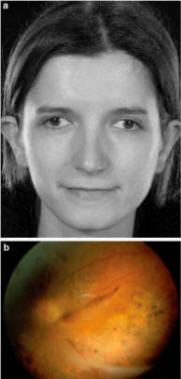
Patients with Wagner syndrome present early in life with pseudostrabismus from congenital temporal displacement of the fovea Klinger-Wegener syndrome

What is Wagner syndrome?
Wagner syndrome is a hereditary disorder that causes progressive vision loss. Loss of vision usually begins in adulthood, although some individuals are affected in adolescence. Vision progressively worsens with age.
In people with Wagner syndrome, the light-sensitive tissue that lines the back of the eye (the retina) becomes thin and may separate from the back of the eye (retinal detachment). The blood vessels within the retina may also be abnormal. Additionally, the thick, clear gel that fills the eyeball (the vitreous) becomes watery and thin (vitreous degeneration). Some people with Wagner syndrome have blurred vision because of ectopic fovea, an abnormality in which the part of the retina responsible for sharp central vision is out of place. Affected individuals may also experience nearsightedness (myopia) or a clouding of the lens of the eye (cataract).
A condition called erosive vitreoretinopathy is very similar to Wagner syndrome. In addition to the signs and symptoms of Wagner syndrome, people with erosive vitreoretinopathy experience progressive night blindness and a narrowing of their field of vision. Recent research indicates that erosive vitreoretinopathy is likely a variant of Wagner syndrome.
How common is Wagner syndrome?
Wagner syndrome is a rare disorder, although its exact prevalence is unknown. Approximately 50 families affected by the disorder have been identified worldwide.
What is the History related to Wagner syndrome?
In 1938 Hans Wagner described 13 members of a Canton Zurich family with a peculiar lesion of the vitreous and retina. Ten additional affected members were observed by Boehringer et al. in 1960 and 5 more by Ricci in 1961. In Holland Jansen in 1962 described 2 families with a total of 39 affected persons. Alexander and Shea in 1965 reported a family. In the last report, characteristic facies (epicanthus, broad sunken nasal bridge, receding chin) was noted. Genu valgum was present in all. In addition to typical changes in the vitreous, retinal detachment occurs in some and cataract is another complication.
Wagner's syndrome has been used as a synonym for Stickler's syndrome. Since there may be more than one type of Wagner syndrome, differentiation from Stickler's syndrome is difficult, and authors disagree as to whether these are the same entity. It may be that Wagner has skeletal effects, but not the joint and hearing problems of Stickler's syndrome. Blair et al. in 1979 concluded that the Stickler and Wagner syndromes are the same disorder. However, retinal detachment, which is a feature of Stickler' syndrome, was not noted in any of the 28 members of the original Swiss family studied by Wagner in 1938 and later by Boehringer in 1960 and Ricci in 1961.
Current Developments
An exhaustive genetics study of blood from 54 patients found everyone with Wagner's disease has the same eight "markers," a genetic fingerprint that sets them apart from those with healthy eyes.
The gene involved helps regulate how the body makes collagen, a sort of chemical glue that holds tissues together in many parts of the body. This particular collagen gene only becomes active in the jelly-like material that fills the eyeball; in Wagner's disease this "vitreous" jelly grabs too tightly to the already weak retina and pulls it away.
Most people with the disease need laser repairs to the retina, and about 60 per cent need further surgery.
Also Known As
Read the case report of wagners disease.
Multifocal neuropathy